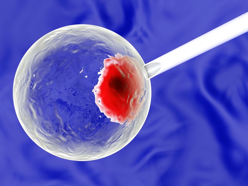
This technology is a unique new laboratory research discovery and diagnostic instrument for multi- mode imaging that has the ability to compare the dynamics of healthy cells vs. diseased cells. A very small Scanning Mass Spectrometry (SMS) probe is inserted into a living cell/ cell culture within an Environmental Scanning Electron Microscope (ESEM) to evaluate the metabolism of the cells over time. ESEM-SMS will enable continuous imaging of the cell’s biochemical activity with a resolution of milliseconds and femtoliters. It will identify metabolic products (biochemicals) indicative of health or disease. Data from this instrument has the potential to bring about a life changing effect on many areas of biomedical sciences.
- Discrimination between healthy & diseased cell biology
- Personalized medicine – specific treatment for an individual
- Diagnosis of a disease prior to symptoms
- Unique capability to sample at the single cell level
This invention would be useful in cancer biomarker discovery, bio-manufacturing process analysis and control, and biosensor development. Additionally, it provides the ability for basic research studies such as determination amyloid aggregation kinetics. Amyloid formation is associated with a number of diseases including Alzheimer’s, Parkinson’s, Huntington’s and Mad Cow Disease.
With the advent of personalized medicine as an emerging trend that utilizes a patient’s genes, proteins and biomarkers to determine medical treatment and diagnosis specific to that patient, tools are needed to move personalized medicine from research to practice. Despite the great interest of using individual data in clinical practice, many obstacles lie in the way of integrating personalized medicine into routine clinical care such as the lack of diagnostic support tools used for decision-making. The ability to understand the change from a state of health to a state of disease at the cellular level is a game changer.

