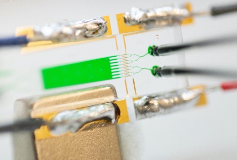
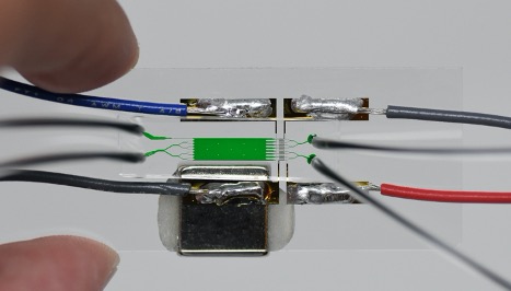
This microfluidic system electronically profiles expression levels of cell surface antigens. It leverages resistive pulse sensing—also known as the Coulter principle—to measure the surface antigen density of cells through magnetophoresis, or the migration of particles in a magnetic field. The Georgia Tech research team has integrated this technology with a microflow cytometry system designed to characterize cells using a straightforward, inexpensive microfluidic device appropriate for point-of-care-settings.
The microflow cytometry system consists of three components: a polydimethylsiloxane (PDMS) microfluidic layer fabricated via soft-lithography, a glass substrate for the sensor network fabricated via lift-off process, and an on-chip permanent magnet. The immunomagnetically labeled cells are driven into a sheath flow and deflected under the external magnetic field according to their magnetic load, which is proportional to their surface expression. The outward flow then divides into channels that utilize a multiplexed sensor network called Microfluidic CODES. Since the various flow conditions can be manipulated, each flow produces unique information about the sample, and high dynamic range can be achieved by collectively analyzing data from various flow rates.
For more information about Microfluidic CODES, see #7107/8032.
- Portable: This innovation uses a disposable, handheld device without bulky or costly equipment, which is expected to be especially useful in low-resource settings.
- Simple: It is designed to achieve results similar to those from a commercial flow cytometer but without requiring initial purification.
- Recoverable samples: Unlike commercial systems, the analyzed sample can be recovered for further tests at the end of the analysis.
- High dynamic range (HDR) operation: Inspired by digital photography, the device can operate in HDR mode by changing the flow rate.
- Health care
- Immunology
- Stem cell transplantation
- Point-of-care diagnosis and monitoring
- Surveillance testing
- Biotechnology
- Biomedical research
- Flow cytometry
Surface antigens are protein complexes on the cell membrane that regulate biochemical interactions of cells. The measurement of their expression levels is widely used in immunophenotyping, clinical diagnosis, and biomedical research, mainly via flow cytometry. Flow cytometers, however, are often complex and costly to operate, which inhibits their use especially in resource-poor settings. This Georgia Tech microfluidic device is designed to serve as a resourceful solution.
To see more technologies by Dr. Sarioglu and his team, please click here.

The working principle of the system

Image of the microflow cytometer

The entire microfluidic chip is as small as a quarter.
