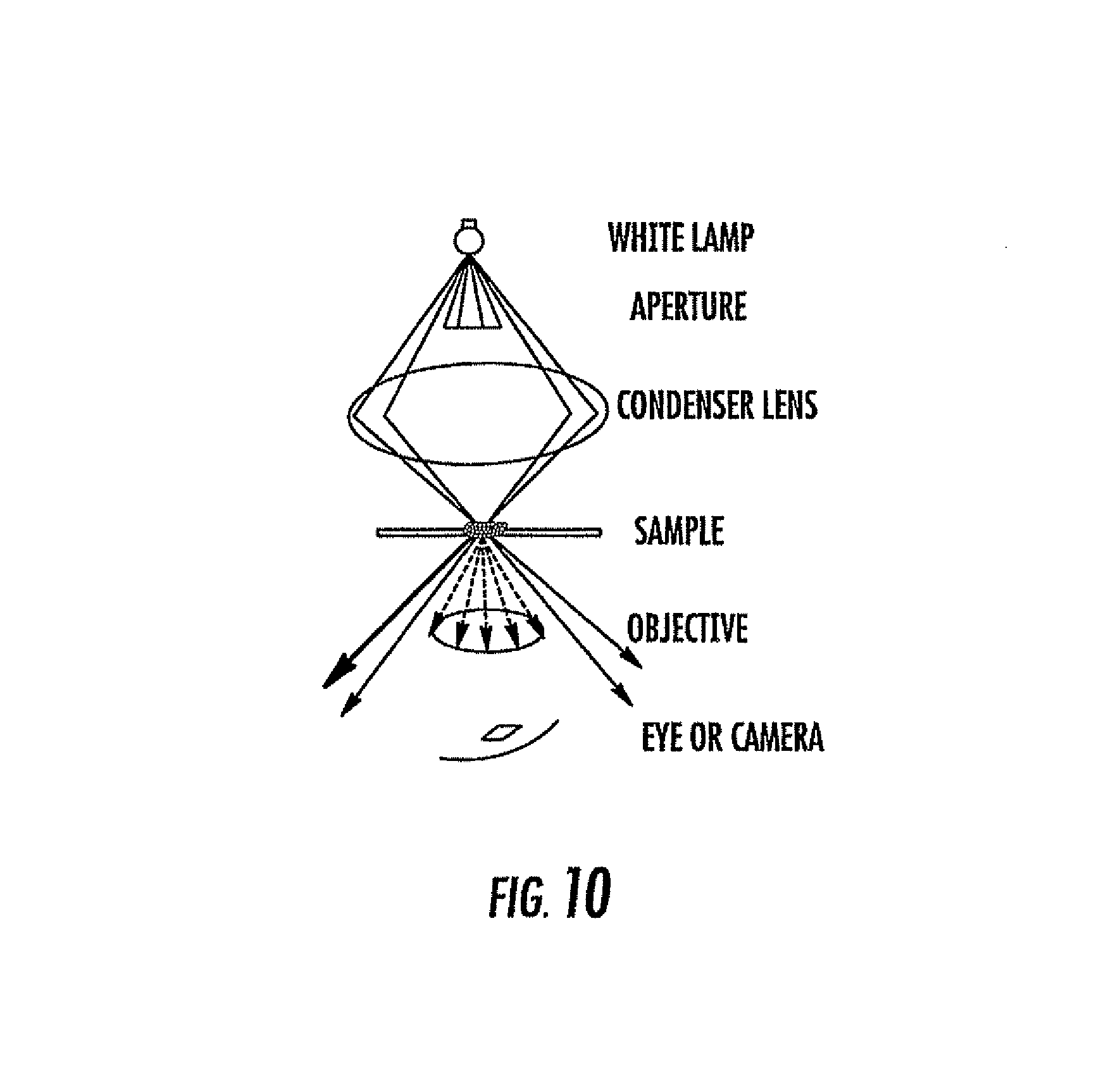
Georgia Tech inventors have found a method that uses nanoparticles of noble metals for molecular imaging, diagnoses, and treatment. Near infrared light is used as a cancer treatment because of its ability for maximum penetration through tissue and to chemically kill malignant cells. The researchers have found a method in which noble metal nanoparticles that have scattering in the near infrared wavelengths can be conjugated to antibodies for use in molecular imaging and diagnoses. This method can be used to differentiate cancer cells from normal cells or to identify the presence of a target substance. They can be differentiated or identified in either living or dead cell cultures (microscope) or in vivo (endoscope) by observing the scattering properties or absorption wavelengths. This involves administering nanoparticles that are conjugated to a specific binding molecule, such as an antibody, to both healthy and malignant cells, and subsequently illuminating them using electromagnetic radiation to compare differences in scattering/absorption patterns between malignant cells and healthy cells. This method provides an improved, alternative approach for molecular imaging and cancer diagnoses and treatment that is easily prepared, non-toxic, and has excellent detecting properties for cellular imaging.
- Can be used with a simple microscope
- More sensitive and cost-effective
- Non-toxic
- Would not harm healthy tissue.
- Easily prepared
- Doctor’s offices
- Clinical labs- cancer detection
- Molecular/cellular imaging field
Cellular imaging techniques are used to provide anatomic detail on the cellular level and are vital for diagnostic applications and research. Current methods include optical probes that use markers attached to antibodies to give information on the presence of specific molecules. Quantum dots are frequently used, but the semiconductor material is potentially toxic to humans, which poses a problem for in vivo applications. The emergence of nanostructures with optical properties, such as gold nanoparticles, have provided new alternatives for diagnostic, therapeutic, and imaging applications. Nanoparticles have been used in photodynamic therapy (PDT) and photothermal therapy for treating cancer in vivo, but have limitations in that heating is too excessive and nonspecific in that destroys both malignant and healthy tissue. Thus, there is a need for improved compositions and methods for detecting or treating different pathologies, including cancer.