Inventors at Georgia Tech disclose a unique new instrument for multi-modal imaging of biological samples with subcellular spatial resolution. The instrument is composed of a combination of Scanning Electron Microscopy (SEM) and a new mode of Desorption Electrospray Ionization (DESI) imaging mass spectrometry. Termed BeamMap, for Beam Enabled Accurate Mapping & Molecular Analyte Profiling, provides untargeted characterization of protein, metabolite and lipid chemistry and correlation with topological features, yielding an order of magnitude improvement in the achievable resolution for electrospray based imaging. Beam Map has potential to bring about a transformative effect on many areas of biomedical sciences.
- Integrative perspective imaging - topological and chemical imaging of complex biological samples and heterogeneous nanomaterial interfaces with sub-micrometric resolution
- Improved spatial resolution - system provides chemical imaging resolution of ~ 250 nm and electron microscopy topological resolution of ~ 50 nm.
- Accuracy in analytics - analysis at the level of a single cell or with subcellular resolution has been seen as a next frontier in innovative approaches to discovery
- Research and Development
- Hypothesis Generator
- Biological and Biochemical Observation
- Molecular Medicine Applications
- Identification of Biomarkers
Multi-mode imaging samples allow for characterization of physical and chemical properties of the samples underlying their functionality for use in applications. Improved spatial resolution in biological and biochemical observation and measurement has been recognized as a key goal in development of instruments and methodologies for health related basic science research, as the richness of data sets and their potential for leading to transformative discoveries are revealed. Recognition of the structural and chemical properties of different molecules serves to assist researchers in areas of molecular separation, data analytics, and data visualization. Ultimately, improving standards of imaging is vital to the mission to improve research tools in biotechnology.

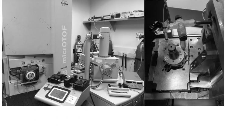
Image shows a mass spectrometer and scanning electronic microscope that provide the foundation for the BeamMap system, which can simultaneously determine surface topology and chemical makeup of a biological sample.

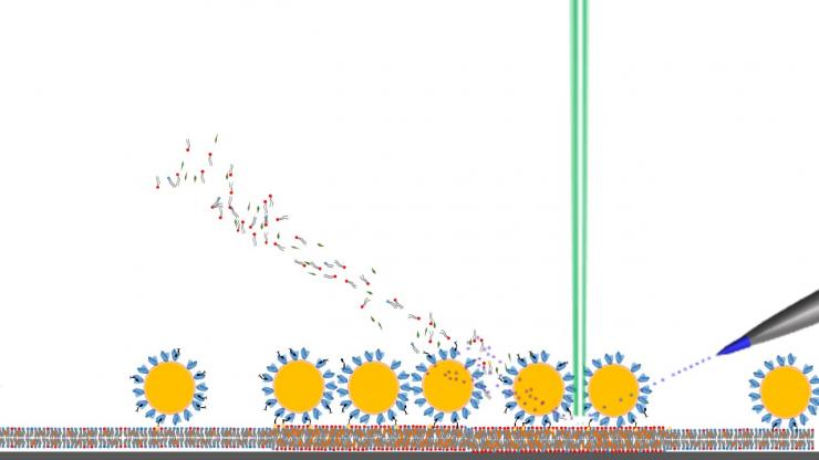
BeamMap uses an electron beam to image topology, provide localized heating to promote desorption and allow precise determination of the location of the spray capillary and electrospray impact area for imaging. The electrosprayed liquid beam at a controlled energy level is used to desorb and charge analytes for chemical imaging in BeamMap.
