*This technology has been exclusively licensed.*
Researchers at Georgia Tech have developed a highly effective, simplified method for hematology analysis using ultraviolet (UV) microscopy. In particular, this new method streamlines a complete blood count (CBC) procedure, the most common form of hematology analysis. This novel technology allows for testing that is critical for diagnosing and monitoring blood and bone marrow conditions to be performed in point-of-care settings, like in the clinic or at home. Deep UV microscopy enables cell analysis without chemical reagents or fluorescent labels that often interfere with image quality and make procedures complex. This technique extracts morphological and molecular features from individual cells and uses machine learning for their classification.
This unique approach quickly collects quantitative endogenous molecular information from live cells without causing any cell damage. The short wavelength of UV light produces high-resolution images that allow for highly specific cell phenotyping which, in turn, leads to more accurate diagnoses and treatments.
- Streamlined: Enables testing in point-of-care settings without complex procedures and specially trained personnel
- Highly accurate: Offers a spatial resolution higher than current methods due to the shorter wavelength of UV light
- Cost-effective: Saves on reagents, complex equipment costs, and time-intensive processes without compromising diagnostic quality
This technology offers an improved and simplified approach for complete blood count analysis that can be used in clinical and diagnostic testing settings. It has applications for the diagnoses and treatment of blood and bone marrow conditions including:
- Anemia
- Hemophilia
- Leukemia
- Thrombocytopenia
- Neutropenia
- Sickle cell disease
- White blood cell counting (five-part differential)
- Infections
- Inflammation
Hematology analysis is a critical tool for the diagnosis and treatment of blood conditions and diseases. Often, however, its methods are costly and laborious, requiring specially trained personnel, complex equipment, and multiple chemical reagents. Many current standard-of-care models evaluate the morphology, population, and molecular or cytogenic properties of blood cells using several different modalities, such as absorption spectroscopy, impedance measurement, and flow cytometry. The UV microscopy hematology analysis method developed by Georgia Tech moves beyond the obstacles of current models while delivering highly accurate and timely results. This means lower costs for doctors, clinics, and caretakers as well as higher quality care for patients.
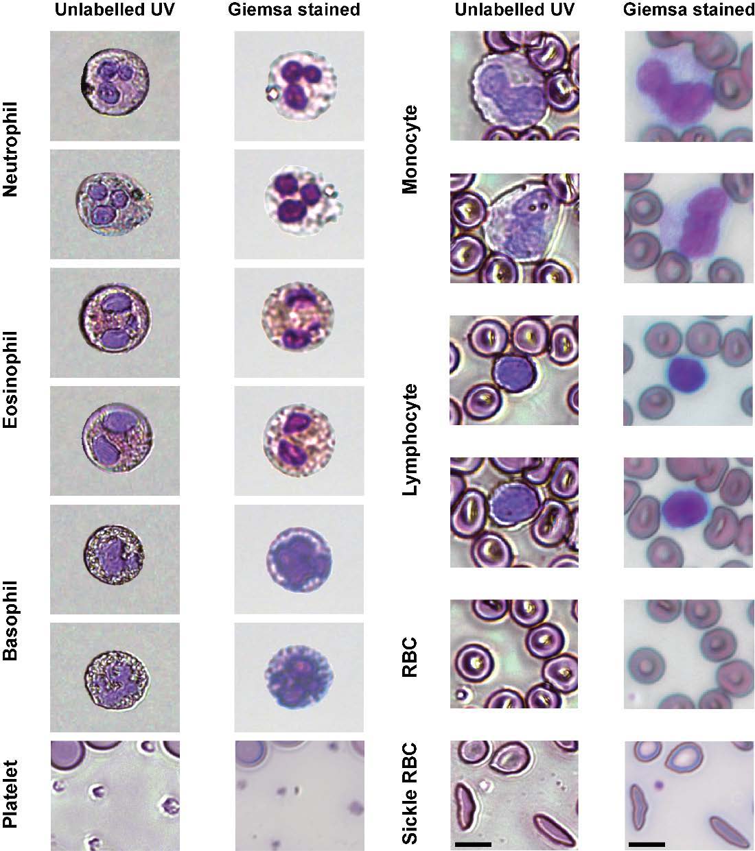
Various blood cell types imaged using the Georgia Tech UV microscopy method compared to images of the same cells stained with a nucleic acid stain