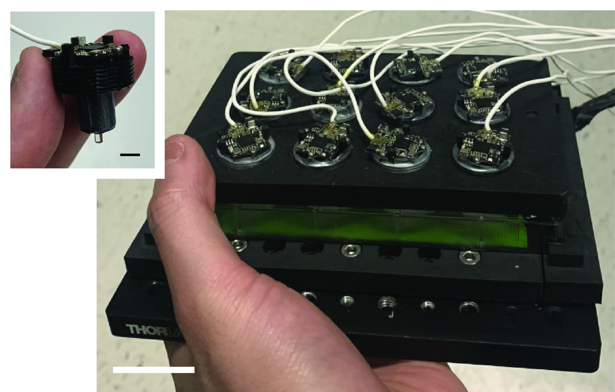
This miniaturized modular-array microscopy (MAM) system enables fluorescence live-cell imaging in flexible and portable formats. The system consists of an imaging device compatible with various in situ conditions and associated with a control algorithm for its operation and data acquisition. The imaging device contains three main components: an array of modular microscopes, a mounting cover, and an illumination panel. The MAM system leverages emerging miniscopy technology that provides numerous benefits in fluorescence imaging capability, flexibility, and scalability.
Georgia Tech researchers present a new upright architecture that integrates gradient-index objectives and illumination and acquisition modules. The modular design allows for feasible parallelization of multi-site data acquisition from conventional well plates.
Georgia Tech’s system provides high fluorescence sensitivity, efficiency, and resolution for capture of spatiotemporal details. It can be applied to various large-scale specimens beyond single cells by implementing motorized axial scanning. The imaging scheme represents minimal instrumental complexity and full compatibility with various cell cultures, conventional microscopy or sensing modalities, and microfluidic configurations. This permits various levels of integration for long-term in situ single-cell imaging and analysis at the chip scale.
The technology has been demonstrated using various biological samples and experimental conditions.
- Powerful: Offers high fluorescence sensitivity, efficiency, and spatiotemporal resolution (~3 µm and up to 60 Hz)
- Configurable: Offers compatibility with conventional cell culture assays and physiological imaging, providing accessibility to upright physiological imaging and integration with biochemical sensors under the cell platform
- Efficient: Provides effective parallelization of multi-site data acquisition
This innovation offers long-term, in situ, time-lapse, imaging of live cells in flexible and portable formats. It is particularly useful for:
- Microscopy
- Bioimaging
- Clinical cell screening
- Drug discovery
- Research
Visualizing diverse anatomical and functional behaviors of single cells provides critical insights into the fundamentals of living organisms. Conventional assays rely on standard cell fixation to observe and characterize cells at discrete intervals and are insufficient to reveal many dynamic, rare, and heterogeneous cellular events.
Fluorescence live-cell imaging allows for continuous interrogation of cellular behaviors, and recent developments in portable live-cell imaging platforms have rapidly transformed conventional schemes with high adaptability, cost-effective functionalities, and enhanced accessibility to cell-based assays. However, broader applications remain restricted due to issues such as compatibility with conventional cell culture workflow and biochemical sensors, accessibility to upright physiological imaging, and parallelization of data acquisition.
Georgia Tech’s MAM system provides high fluorescence efficiency and resolution and enables parallel data acquisition using conventional off-the-shelf cell chambers, offering a potentially promising advancement to single-cell imaging and analysis.

Photograph of the fully assembled MAM system, with an inset photograph showing a single modular detection unit
