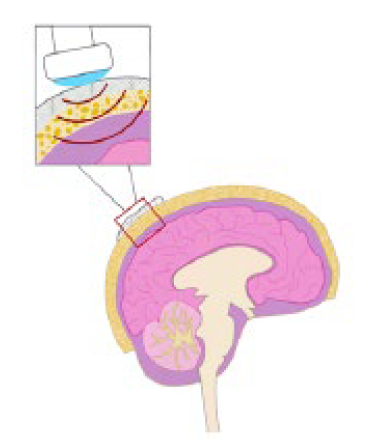
This novel method may expand the application of ultrasound treatments to the brain by allowing ultrasound energy to penetrate the skull for research and therapeutic purposes. It uses a non-Hermitian complementary metamaterial (NHCMM) that helps counteract the impedance mismatch and energy attenuation effects of the skull. These effects typically prevent transmission of high-frequency ultrasound and have previously limited the use of ultrasound for imaging and treatments for the brain.
Georgia Tech’s method has been shown to achieve near-perfect signal and energy transmission through the skull at high frequencies, which may lay the foundation for noninvasive ultrasonic brain imaging, stimulations for in-vivo brain circuitry research, and treatments for neural disorders including brain tumors and strokes—all with the skull fully intact. In addition to the convenience of portability, Georgia Tech’s method may prove to be a less expensive diagnostic modality compared with MRI, CT, and other imaging techniques.
- Portable: Designed to enable imaging of the brain via small, mobile ultrasound devices, which may offer greater convenience compared with MRI and CT machines
- Affordable: May be a lower cost modality compared with leading techniques, such as MRI, CT, or positron emission tomography (PET)
- Robust: Demonstrates energy transmission through the skull on par with that of an aqueous medium in preliminary testing
- Non-invasive: Designed to enable imaging, diagnosis, and treatment of the brain with the skull fully intact without exposing patients to high doses of radiation
Georgia Tech’s innovation may be broadly applicable to hospitals, medical research institutions, and health care facilities, particularly those engaged in:
- Non-invasive ultrasound imaging
- In-vivo brain circuitry research
- Treatments for neural and brain disorders
- Evaluation of strokes
- Identification of brain tumors and lesions
- Enhancing drug delivery through the blood-brain barrier
Neurological disorders are the leading cause of disability and the second leading cause of death worldwide. With more than a million people in the U.S. alone diagnosed with these conditions each year, it is no surprise that treatment advances are a subject of great interest. While brain imaging technologies such as MRI, CT, and PET are valuable for understanding these disorders as well as for diagnosing and treating them, they may not be suitable for all clinical applications given their expense, large size, lack of portability, and radiation risks for patients.
Ultrasound offers a portable, affordable, and safe alternative, but its application has been limited because it does not travel well through air and bone. For brain disorders, it is typically only used in infants, whose soft spot provides an acoustic window for imaging the skull, and rarely in adults who have had parts of their skull removed from surgery or injury.
Georgia Tech’s method is designed to allow ultrasound to penetrate the skull and may thereby open the door to new possibilities for brain imaging and treatment, including disease diagnosis, brain disorder treatment, stroke evaluation, and brain tumor identification.

This diagram shows an ultrasound probe and Georgia Tech’s non-Hermitian complementary metamaterial.
