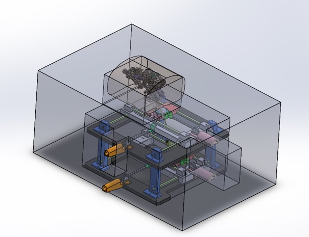
This actuator-guided positioning and control system enhances magnetic resonance elastography (MRE) for precision-guided diagnostics and interventional treatments. Used in magnetic resonance imaging (MRI) applications, the system incorporates an extremely precise positioning mechanism capable of sub-millimeter motions within the MR environment, a high-frequency MRE actuator, and an imaging and control scheme that provides position control and image analysis. This system allows movement of the actuator relative to a region of interest within the bore of an MRI scanner and can create 3D images in near real time to guide procedures.
The Georgia Tech system works with a five degrees of freedom (5DOF) MRI-compatible robot that uses visual servoing to reach a targeted contact and orientation. Accurate positioning of the shear wave fields relative to the area of study is critical and is optimized through position control of MRI-conditional piezoelectric direct drive actuators. The parallel plane mechanism enables accurate use of computer vision data to control the motion of the robot with fiducial markers. A software interface enables visualization of MR images.
The device can be used with an MRI scanner as a non-invasive diagnostic tool for soft tissue characterization through the mechanical property of complex shear modulus/stiffness. For example, in spinal column imaging, this device can be used to understand the severity of intervertebral disc degeneration.
- Precise: Extremely precise positioning mechanism enables orientation and control capable of sub-millimeter motions within the MR environment
- Rapid: Significantly speeds up procedure time over standard MRI and offers wavefield images in near real time
- High resolution: Uses high-frequency actuation to enable imaging of small, geometrically difficult targets
- Low cost: Manufacturing costs would be low
- Diagnosis of soft tissues
- Treatment and diagnosis of spinal column conditions
- Imaging for diagnosis of intervertebral disc (IVD) degeneration
- Intraspinal injections
- Other MRI and MRE applications benefiting from precise and rapid guided positioning of instruments and interventional tools
MRE combines MRI with low-frequency vibrations to create an elastogram, which is a visual map showing stiffness of body tissues. MRE is typically used to measure the stiffness of liver tissue in people with known or suspected liver disease. Georgia Tech’s innovation enhances MRE to enable its use in detecting degenerative disc disease, providing a non-invasive method of diagnosing it in early stages. It also provides precise and rapid position control for intraspinal injections.
The technology is applicable to any MRI and MRE application benefiting from precise and rapid guided positioning of instruments and interventional tools.

Proposed validation setup for the 5DOF MRE actuator positioning system, tissue mimicking phantom fixed above the MRE actuator, with both the robot and phantom set into a low-density foam block.
