Live, 3D, noninvasive observation of intact organoid architectures is now possible with Georgia Tech’s Fourier light-field microscope that utilizes a hybrid point-spread function (hPSF-FLFM). This new microscope system transforms conventional 3D microscopy, enabling exploration of less accessible, spatiotemporally challenging regimes in organoid research. The system offers cellular level (2-3 μm and 5-6 μm in x-y and z, respectively) and millisecond-scale spatiotemporal characterization of whole-organoid dynamic changes that span large imaging volumes (>900 μm × 900 μm × 200 μm in x, y, z, respectively).
Comprising a standard epifluorescence microscope and a light-field acquisition module, the hPSF-FLFM system provides scan-free, fast snapshot recording of intact organoids and their dynamic cellular processes with minimum photodamage per volumetric acquisition compared to existing techniques.
- Cost-efficient and scalable: Is fully adaptable to epifluorescence protocols resulting in a system that is both cost-efficient and highly scalable
- Fast: Captures fast cellular and tissue dynamic processes in a simultaneous, volumetric manner (e.g., collective cellular responses at sub-second time scales across whole samples)
- Validated: Responses to extracellular physical cues such as osmotic and mechanical stresses have been recorded using human induced pluripotent stem cell-derived colon organoids (hCOs).
- Minimizes damage: Provides scan-free, fast snapshot recording with minimum photodamage, unlike conventional imaging methods with underlying scanning mechanism that compromise the time resolution of volumetric acquisition and result in increased photodamage and inability to capture fast cellular and tissue dynamic processes
- Versatile: Images biological systems such as cells and/or tissue of plants and animals, including embryonic stem cells or induced pluripotent stem cells, as well as many types of organoids
- Robust: Facilitates the 3D characterization of cellular dynamic processes of whole organoids in response to rapid extracellular physical cues
- Expandable: Combines versatile optical and computational strategies to enable the hPSF-FLFM system to potentially to expand beyond organoids to in vivo organ research
This technology advances volumetric interrogation of organoid systems as a research tool for:
- Modeling tissue development and disease
- Biomaterials and protocols
- Drug discovery
- Provides critical insights for understanding:
- Regenerative medicine
- Tissue homeostasis
- Organ function

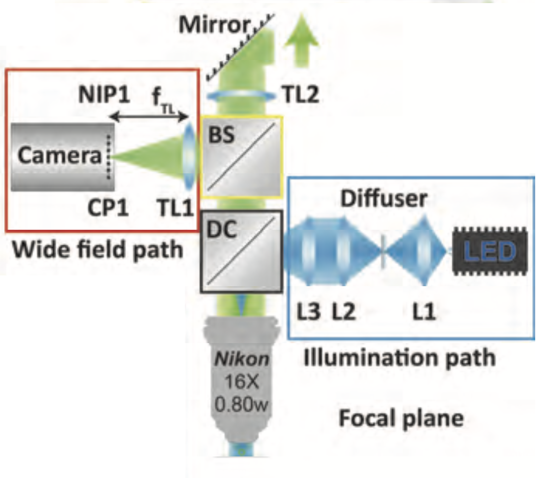
Schematic of the setup. Camera plane (CP); tube lens (TL); diachronic cube (DC); beam splitter (BS); L1, L2, L3 lens to homogenize and focus the LED light onto the back focal plane of the objective lens; native image plane (NIP); focal length of the corresponding tube lens (fTL)

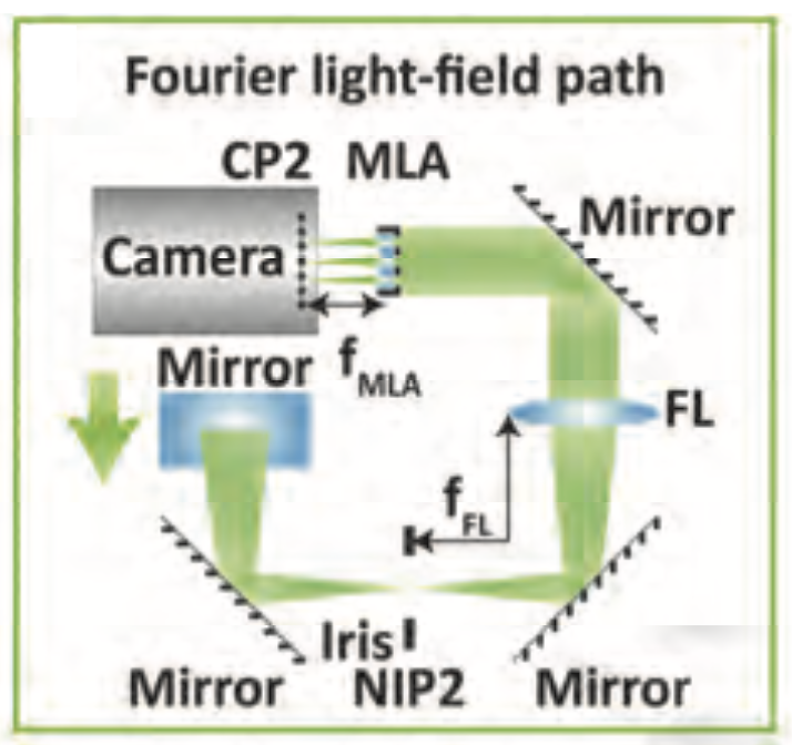
Schematic of the light-field acquisition: Camera plane (CP); Fourier lens (FL); native image plane (NIP); focal length of the Fourier lens (fFL); focal length of the microlens array (MLA) (fMLA)

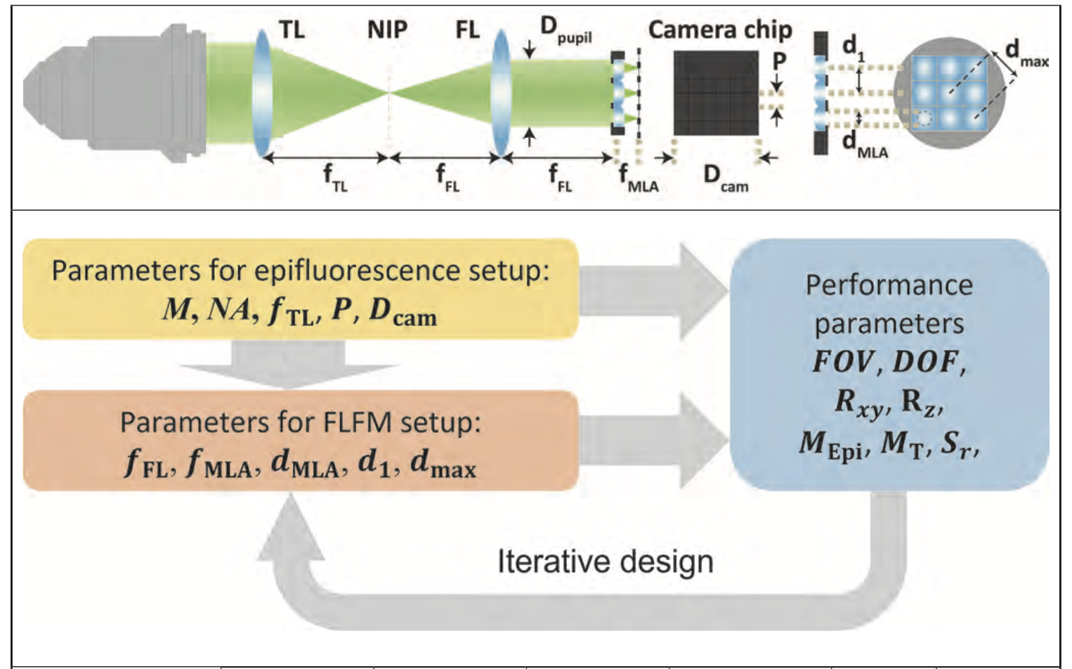
Summary of design and performance parameters of the system
