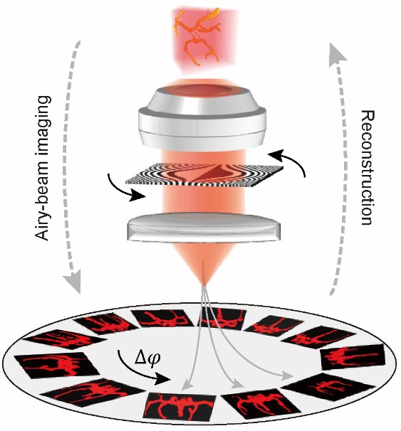
Inventors at Georgia Tech have developed an Airy-beam tomographic microscopy (ATM) technique that allows for scan-free, volumetric 3D cell and tissue imaging using nondiffracting, self-accelerating Airy beams. Conventional wide-field imaging techniques are telecentric, producing orthographic views that require scanning in order to acquire 3D information. In contrast, the self-accelerating propagation trajectory of an Airy beam innately forms a perspective view of the object. Manipulating the Airy trajectories can provide sufficient perspective views to enable the entire volume to be computationally synthesized in a tomographic manner (see the image below). Exploiting highly adjustable Airy trajectories in the 3D space, ATM transforms the conventional imaging scheme in wide-field microscopy, producing a consistent, near diffraction-limited 3D resolution across a ten-fold extended imaging depth. Such a strategy can be readily translated to other non-optical waveforms such as acoustic, plasmonic, and electron waves. The ATM system is potentially applicable to a wide range of biological specimens, spanning molecular, cellular, and tissue levels, thereby offering a promising paradigm for 3D optical microscopy.
- Streamlined: Offers a scan-free method of obtaining 3D-resolution images
- Volumetric imaging: Generates and resolves images of tissues and cells in the 3D space
- High resolution and image depth: Provides 3D diffraction-limited resolution
- High image depth: Produces a tenfold improvement in image depth compared with conventional wide-field biological microscopy
- Optical microscopy for biological imaging
- Optical manipulation for microfluidics applications in biological sciences
- Laser filamentation for applied physics in remote spectroscopy
- Micro-machining for myriad applications from biomedical to MEMS
- Non-linear optics for myriad applications from computers to sensors
- Generation of varying non-diffracting waveforms (i.e., electron beams)
- Generation of plasmonic waves (biochemical sensors, and others), acoustic waves (electronics), and quantum particles
Airy-beam-enabled optical imaging has great potential to advance biological research and clinical diagnoses, enabling precise 3D localization of single molecules in super-resolution microscopy as well as enhanced field of view and image quality in light-sheet microscopy. Many biological sciences fields stand to benefit, including regenerative medicine, cancer biology, neuroscience, developmental biology, and more. However, these methods primarily implement Airy beams as static point-spread functions of the microscopy systems, rather than exploiting the full capability of the adjustable Airy trajectories. By contrast, Georgia Tech’s method explores the entire 3D space for volumetric imaging of biological specimens. The method also takes advantage of the non-diffraction of Airy beams to effectively mitigate the trade-off between the depth of focus (DOF) and the lateral beam diffraction, thereby offering a depth-invariant resolution across a substantially improved DOF.

Principle of ATM. The object is imaged with Airy beams of varying azimuthal angles of ∆φ and tomographically reconstructed using the recorded perspective images.
