Achieve fast, single-shot, 3D, multi-color endoscopic imaging that detects feature depth with sub-millimeter accuracy and enables quantitative volumetric 3D renderings of tissue. Georgia Tech’s new technology uses a hexagonal gradient index (GRIN) lens array. The GRIN lens array microendoscopy (GLAM) system is designed with a small-diameter stainless steel shaft, built-in illumination, and multi-color imaging. With its combination of optical functionality and realistic endoscope design, GLAM enables GRIN lenses to be practically applied to rigid endoscopy, paving the way for future development using this optical paradigm. The system achieves 3D resolution of ∼100 µm over an imaging depth of ∼22 mm and a field of view up to 1 cm2.
GLAM has the potential to increase safety and efficiency in complex endoscopic procedures as well as improving instrument control for robotic surgery.
- Enhanced control and safety: Real time visual navigation enhances guidance and intervention in complex microenvironments, maintaining high spatial resolution while providing quantitative depth information
- Comfortable: System does not require 3D glasses that can cause physician dizziness or headaches after long periods of use
- Streamlined: Technique eliminates the need for other imaging procedures such as micro-CT or MRI to quantify the 3D morphology of tissue
- Accurate depth: Simple chromatic calibration provides accurate RGB depth encoding and enables quantitative depth estimation
- Fast readout: Pinhole image offsets in the off-axis elemental images provide a quick readout for axial chromatic aberrations in the GRIN lens array, offering fast readout of axial aberrations suitable for incorporating into the downstream analysis
- Adaptable: Small form factor aligns with typical clinical design, making the system readily translatable to a clinical prototype
- Increased safety and efficiency for procedures related to:
- Clinical screening
- Diagnostics
- Interventional treatment (particularly in surgical areas with in-depth extension, vascular encasement, or dense tumor structures)
- Enhanced medical training
- Improved instrument control for robotic surgery

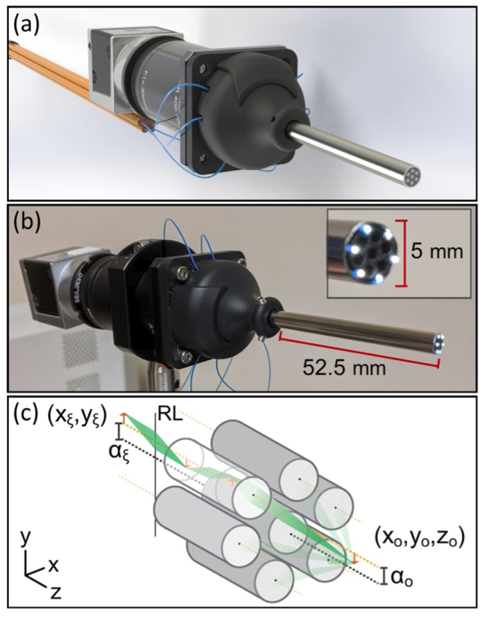
GRIN lens array microendoscopy design and image formation. (b) Assembled endoscope system with overlaid dimensions of the insertion tube. Inset shows a close-up of the 3D printed core of the insertion tube that holds the seven GRIN lenses and six illumination fibers (c) Schematic diagram of light propagation through the system. RL, doublet relay lenses

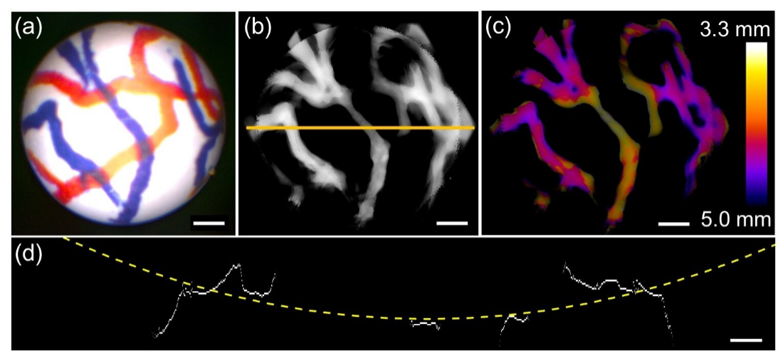
Imaging phantom curvature. (a) Raw image from center endoscope lens of phantom blood vessels wrapped around a 0.5-inch diameter cylinder. (b) Full-color reconstruction of the phantom target, shown in an inverted grayscale image. (c) Depth-coded reconstruction in (b) with distance from the endoscope shown by a color gradient. (d) Projected top view of the reconstruction stack along the yellow line in (b). The profile was fitted with a dashed circle with a diameter of 0.546 inches. Scale bars: 1 mm (a-c). 500 µm (d).
