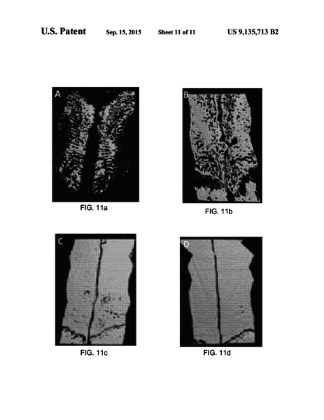
Christopher Hermann from the School of Biomedical Engineering at Georgia Tech has developed an active contour modeling (‘snake’) algorithm that can be used to segment images produced by computed tomography (CT) or microcomputed tomography. The program provides an automated methodology to identify the images of bones, estimate the preliminary boundary areas, and subsequently process the image using a snake algorithm to refine and optimize the image to clarify bone boundaries.
- Increased accuracy: Computer processing removes the need to manually interpret boundaries
- Fast: The algorithm is extremely rapid, repeatable, and accurate
- Versatile: Programs can be implemented in assembly or machine language
- 3D imaging: Images of bones and structures are reconstructed in three dimensions
- Greyscale images
- Traditional & robotic surgery
- Analysis of the growth plate
- Fracture healing
- Studying density changes associated with osteoporosis
This technology was developed to provide a fast, easy, and accurate method to process medical images. Accurate interpretation of medical images, which are typically greyscale, requires the ability to differentiate the boundaries between various tissues, such as bone, vasculature, and organs. Although these different types of tissues can be identified by variations in their values, many medical procedures require a level of precision that is not possible with manual interpretation of images.