This non-invasive, wearable technology tracks in real time the post-injury health of ankle joints. Georgia Tech’s new device allows patients and clinicians to monitor the health of the joint between visits and during activity, providing quantitative data about joint swelling and soft tissue integrity.
Using bioimpedance analysis—a non-invasive method for assessing the composition of tissue—Georgia Tech’s low-power wearable device is designed to enable an accurate and robust assessment of edema and tissue damage that may be caused by excessive activity. The system also uses an inertial measurement unit (IMU) along with bioimpedance signals to evaluate and detect edema and muscle tears, which are crucial for physicians to understand the recovery progress and the patient’s ability to return to activity.
Data from this technology allows users to make better decisions about their activity levels and enables clinicians to monitor and better tailor a rehabilitation course for them. Unlike current joint monitoring technologies, this system can assess ankle health without the need for another device on the healthy contralateral joint.
- Better decisions: Helps clinicians more easily determine the best course for ankle rehabilitation and improves compliance with physical therapy
- Faster recovery: Enables improved rehabilitation guidance based on collected telehealth data that may speed the time to recovery
- Fewer re-injuries: Supports clinicians in making better decisions regarding appropriate activity levels and readiness to return to activities to avoid re-injury
- Easy to use: Tracks joint edema without interfering with the patient’s normal daily activities
- Orthopedic clinics and clinicians
- Professional and recreational athletes
- Physical therapy clinics
- Fitness and recreation centers
- Workplaces requiring standing for long periods
It is estimated that 23,000 Americans sprain their ankle every day, making it the most common musculoskeletal injury. Recovery involves many stages of physical therapy designed to restore function, improve flexibility, and rebuild lost strength in the affected joint. This technology facilitates the tracking of edema in a recently injured ankle joint, specifically acute edema caused by repetitive stress. Tracking acute edema is often challenging for medical professionals as it is induced by certain activities that are difficult to perform in the clinic, such as higher intensity workouts.
How It Works
Extracellular fluid associated with ankle edema freely moves in the joint space as the position of the ankle changes. For example, as a person rotates their ankle, the edema moves around inside the joint space, influenced by gravity and the forces exerted by the structures inside and around the ankle. Electrodes positioned proximally and distally to the joint for bioimpedance measurement inject a low current into the ankle. Different frequency components of this current travel at different depths within the tissue, with low frequency current flowing deeper into the tissue since it cannot penetrate cell membranes and must travel along extracellular paths. In contrast, higher frequency current can penetrate cells, so it travels along a more superficial, shorter path between the electrodes.
This technique leverages the frequency dependency of bioimpedance, comparing the range of reactance at 5 kilohertz to the range of reactance at 100 kilohertz. It then interprets the measurements based on a quantitative simulation model of the effects on joint bioimpedance of variations in edematous fluid volume, muscle fiber tears, and blood flow changes. An IMU is incorporated into the wearable device to capture the angular velocity of the foot, which is used to determine if the subject is moving. It allows position and movement of the limb to be measured during the gait cycle.

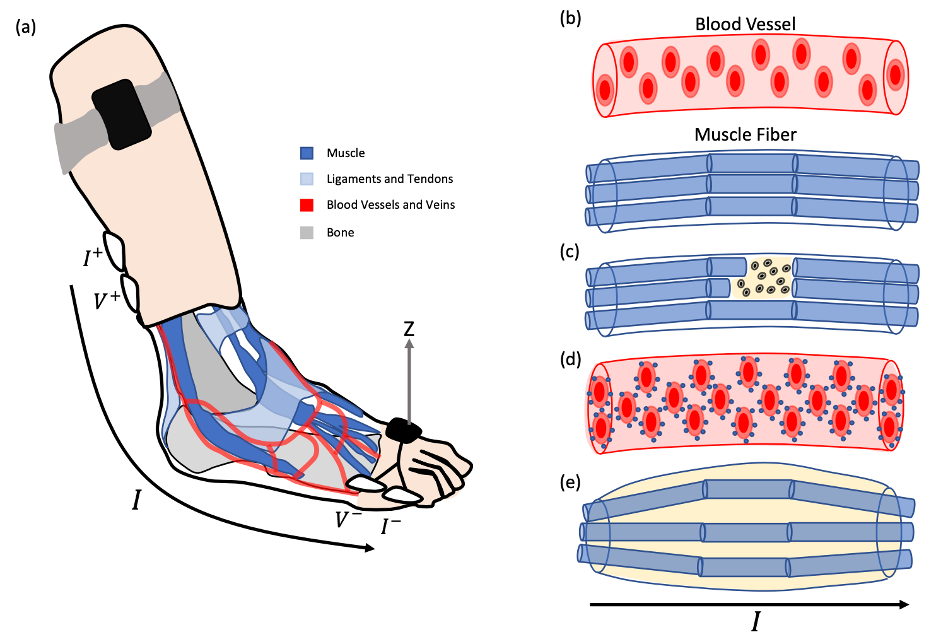
a) The anatomy of the ankle joint showing the lack of fatty tissues or static liquids
b) A sample blood vessel and muscle fiber, which are the primary path for the current applied to measure the
bioimpedance
c) A muscle fiber tear showing the migration of intracellular fluids to the extracellular space surrounding it
d) An increase in the red blood cell count and glucose due to sustained muscle activity
e) An increase in edema due to muscle inflammation

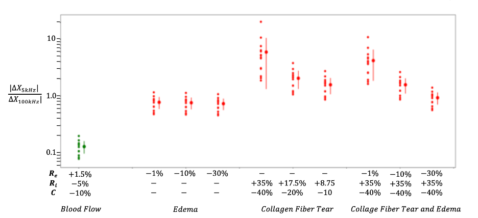
Logarithmic scale of the ratio of the change in the low-frequency reactance to the high-frequency reactance due to simulating the effect of edema, a collagen fiber tear, and a collagen tear accompanied with edema on the baseline ankle impedance of 14 healthy subjects
